


ZRT Webinar: Before & After Cryoablation Images
Ingrid was a guest lecturer for ZRT Laboratory and conducted a one hour webinar on May 26, 2016 for 1,200 providers and women nationally.
Ingrid spoke on her research regarding environmental estrogens / xenostrogen exposure, how it increases your breast cancer risk or risk of reoccurrence and what you can do to limit that risk through her Proactive Breast Wellness Program.
A really exciting part of the webinar was Ingrid’s introduction to “Breast Cryoablation: the new future in breast cancer treatment without surgery.” She presented her Infrared slides of a woman she saw pre and post-cryoablation.
Ingrid was invited to present her case study slides of Mary L. in China at the 5th International Cryoablation Conference at Fuda Hospital outside Canton, China, to 200 researchers. Laura Ross-Paul presented the slides on July 2, 2016 at Fuda Hospital. Ingrid and Laura created the “Early Freeze Protocol,” which was part of the presentation to the international researchers.
Laura Ross-Paul received her cryotherapy treatment in 2003 at the Karmanos Cancer Center in Detroit, Michigan. Laura Ross-Paul of Portland, Oregon, calls herself a “patient pioneer,” as the first woman in the world to receive cryoablation by Dr Peter Littrup as the primary treatment for her multi-focused breast cancer.
Before and After Imagery
Ingrid Edstrom was invited to present her slides and research at the 5th International Forum on Cancer, Cryotherapy and Immunotherapy at Fuda Hospital in the city of Guangzhou (formally Canton) to over 200 physicians and international researchers.
Laura Ross-Paul presented Ingrid’s slides on July 2, 2016, along with the “Early Freeze Protocol” which Edstrom and Paul co-created. We have taken the images from the presentation and displayed them below to further illustrate the before and after effects of cryoablation treatment.
Mary L: Infiltrating Ductal Carcinoma
3 Months Pre-Cryoablation
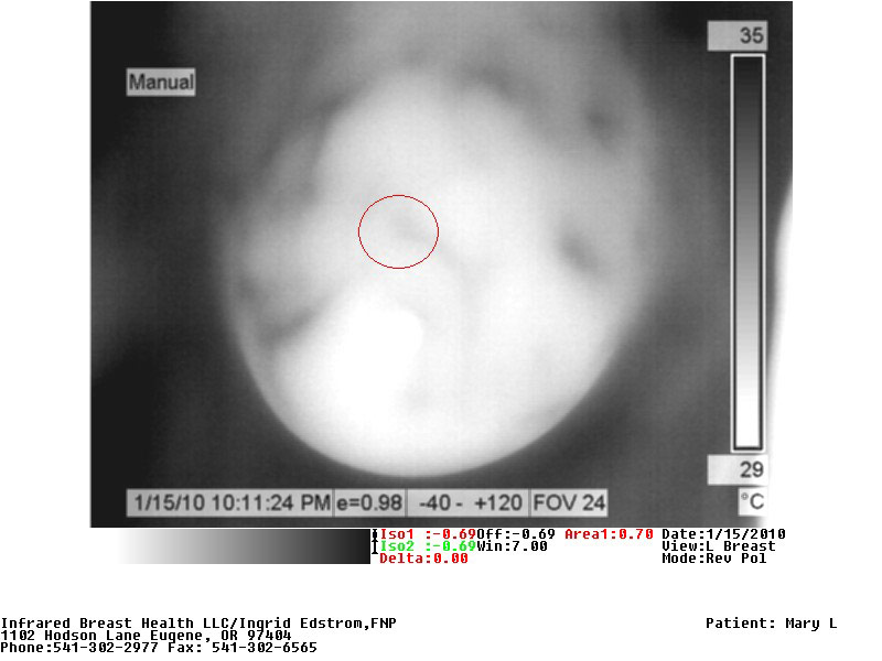

Mary was diagnosed with infiltrating ductal Stage II carcinoma 2 cm by 2 cm tumor in her left breast with dimpling at 12:00. Mary had a very abnormal thermogram with a 2 degree Celsius Delta thermal shift over the mass and blood flow leading to the tumor. The red circle surrounds the region of the tumor. Note there is an enlarged vessel leading to and feeding the cancer visible on the grayscale image (click to enlarge images).
Please note the gray and colored images are of the same view of the breast. The temperature scale on the right of the images, measured in degrees Celsius. On the color images, cold is denoted in dark blue and turns red as the temperature increases. On the grayscale, black is hot with greater metabolic activity. Blood vessels are more visible in the grayscale images.
8 Months Post-Cryoablation
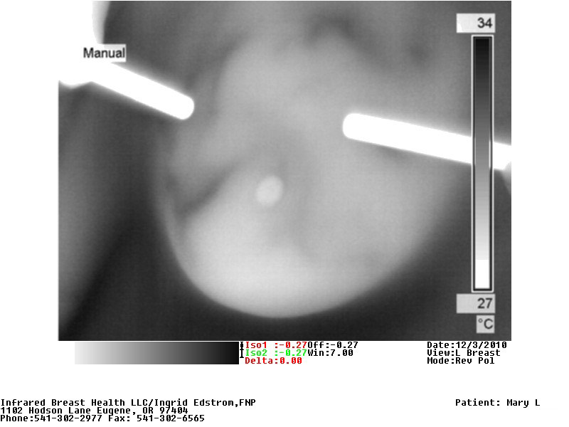
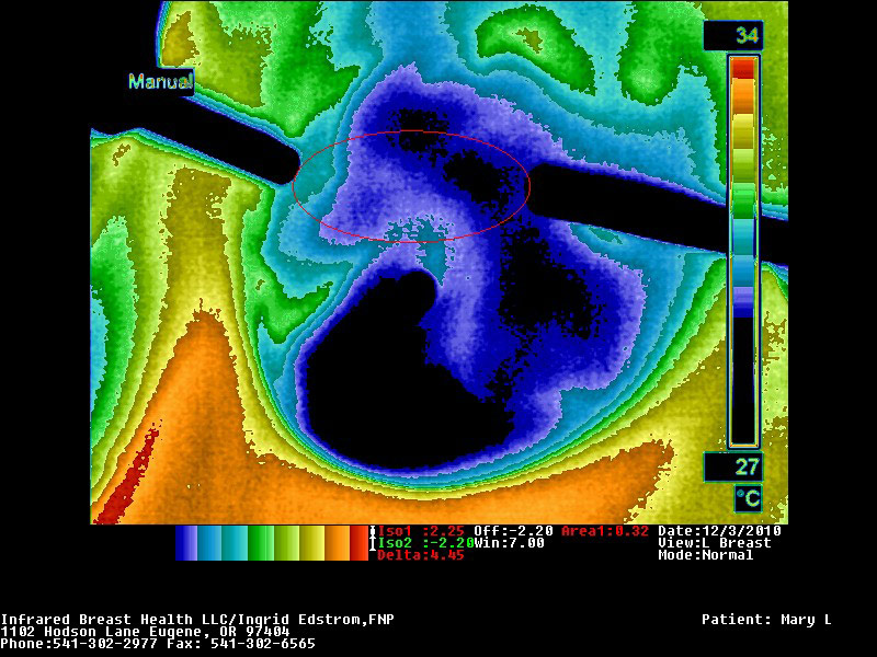
Mary returned 8 months post cryo and now has a large/spongy mass which currently measures 4 cm x 6.5 cm. Previously, the tumor, which was a hard mass, measured 2cm x 2cm. There was no thermal activity over the soft mass and the blood vessels that were observed 3 months prior to her cryotherapy on IR were now absent. The spongy mass represents the “immune effect” that occurs with cryoablation.
Infiltrating ductal carcinomas as well as lobular cancers, when the tumors achieve a certain size, the cancer cells start emitting nitric oxide which is a vaso dilator that keeps the blood vessels open so tumors have greater access to oxygen and nutrition which encourages tumor doubling times. You can see these vascular changes on my website in “Save Your Breasts”
I was alarmed by the size of the soft spongy mass but Dr Littrup told me that this was common after cryotherapy due to the “Immune effect” which occurs in about 85% of the time in the woman’s own body. The body’s immune system is able to now recognize the protein structure of the cancer cells and the body sends out white blood cells and cytokines to clean out the dead tissue that the liquid nitrogen had frozen and this naturally creates immunity to the cancer. You can see the pointer views of the size of the mass on the slide.
NEVER surgically disturb or needle biopsy the spongy area which is comprised of dead tissue and white cells. Biopsy interferes with your own body’s immune system to break down the dead cancer cells and might cause a life threatening infection. Let your body do the healing.
2 Years Post-Cryoablation

Two years post cryotherapy there was a 1.5 cm thermally inactive “cold” mass of scar tissue without any blood flow seen on the slide. Dr Littrup told me, the spongy mass shrinks about 80% in size leaving a small bit of scar tissue. Mary was also instructed about my Proactive Breast Wellness protocol. The red circle surrounds the scar tissue at 12 o’clock.
Video:
Please watch the video Laura Ross-Paul and Ingrid created of the “Early Freeze Protocol” presented at the 5th International Cryoablation Conference at Fuda Hospital outside Canton, China, to 200 researchers.